MCS31 Live Cell Imaging System
MCS31 Live Cell Imaging System - 20x is backordered and will ship as soon as it is back in stock.
Protect your product
Protect your product
Select from a range of extended warranty and service plan options, so you can enjoy your product stress-free. Contact us for more details.
Delivery and Shipping
Delivery and Shipping
We provide delivery solutions tailored to each order to ensure safe and timely arrival of your equipment. Please contact us for more available options
Description
Description
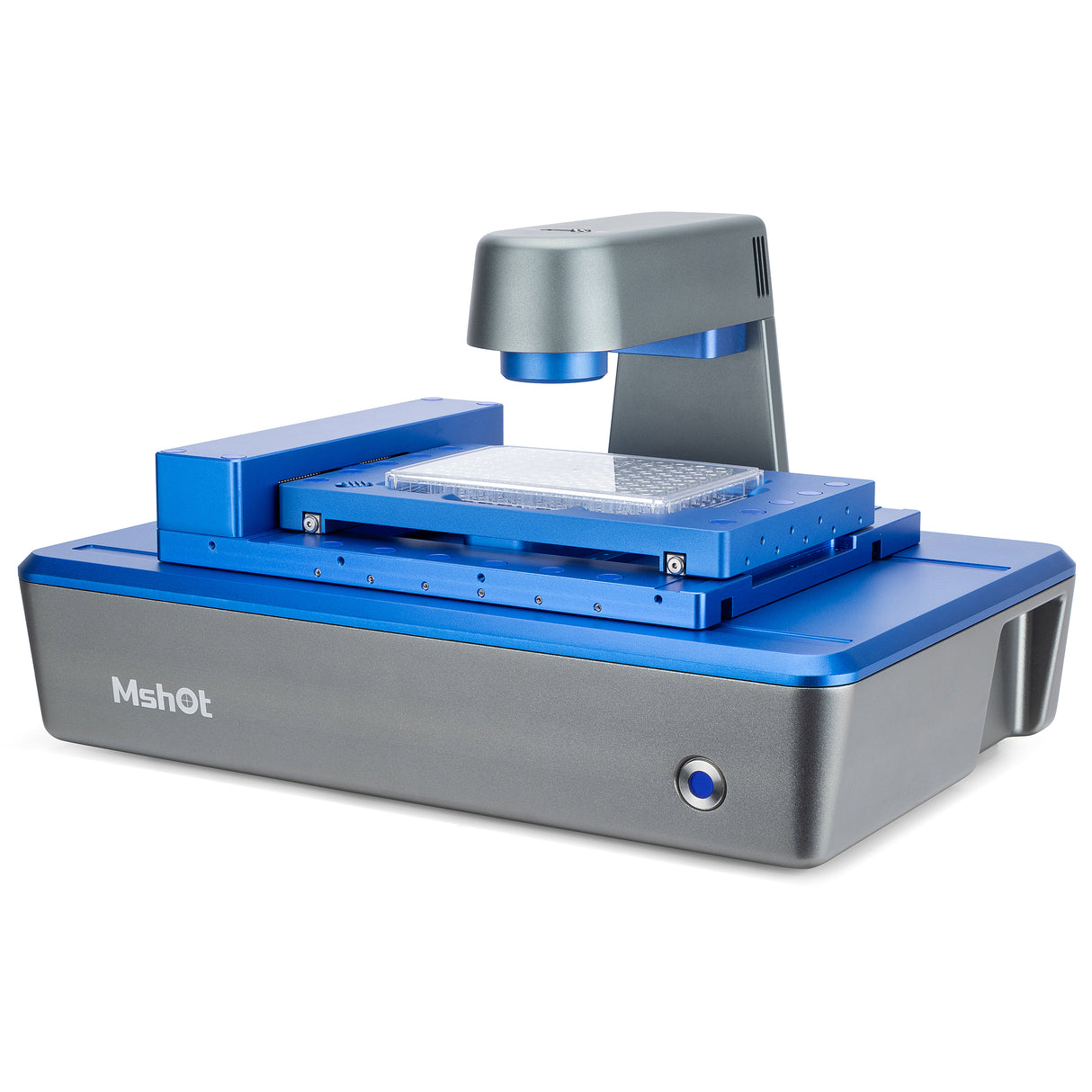
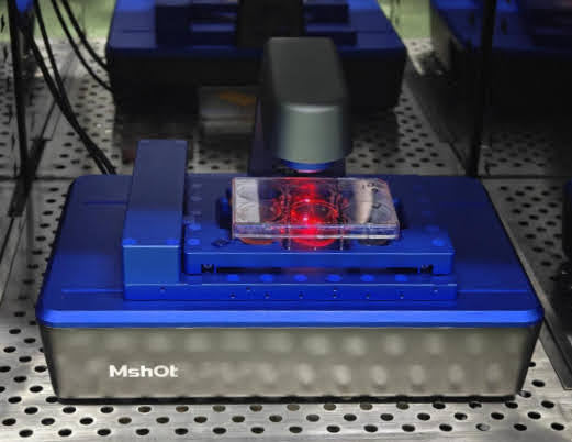
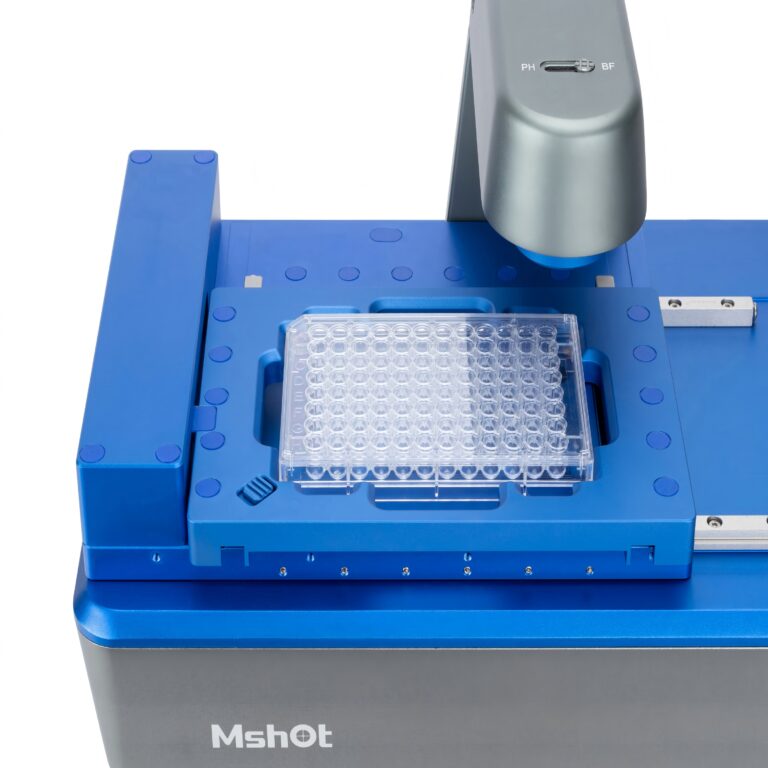
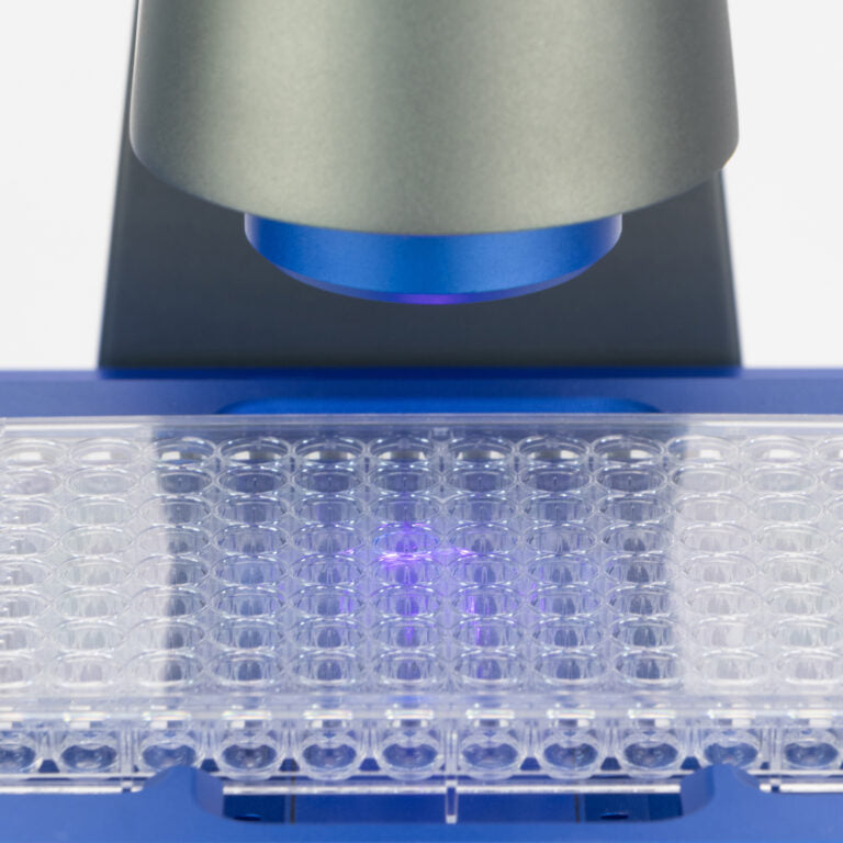
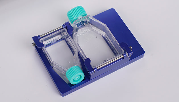
MCS31 is an excellent companion for cell culture. It can be used inside the incubator and supports bright field, phase contrast and fluorescence observation of various culture flasks, petri dishes, as well as 6-well to 384-well plates. It enables scanning and stitching of selected regions, allowing for large-field-of-view, long-term observation and recording of multi-well plates. This significantly improves experimental efficiency in application scenarios such as comparative experiments and reduces the risk of experimental failure caused by contamination and other factors.
| Specifications: | |
| Transmitted Observation | Transmitted bright field / phase contrast illumination, working distance 55mm |
| 50,000 – hour long lifespan, 625nm low – phototoxicity LED light source | |
| Fluorescence Observation (Wavelengths Customizable) |
Blue (B): 472 – 495nm |
| Green (G): 543 – 560nm | |
| Ultraviolet (UV): 393 – 416nm | |
| Objective Lenses | Select any two from 4×/10×/20× objective lenses; Motorized objective lens switching |
| XY Stage | Motorized XY moving platform with a stroke of 115*77mm; Repeat positioning accuracy ≤ 3μm |
| Z Axis | Motorized with auto-focus mechanism |
| Camera |
High speed and high sensitivity camera, 5M 2/3 inch, 40fps |
| Software | Support remote control, including light control, camera control, single image and time lapse acquisition |
| Analysis Function | Cell counting, cell confluency, scratch assay, transfection efficiency, viability analysis |
| Scanning Function | - Multi well imaging and multi position imaging with bright field, phase contrast & multi-channel fluorescence overlay |
| - Multi area scanning, imaging and stitching with bright field, phase contrast & multi-channel fluorescence overlay | |
|
Data Transmission and Power Supply |
USB data cable + DC power cable |
| Working Environment | 5 – 40°C, 20 – 95% RH |
| Dimensions | 252mm × 377mm × 208mm |

